Anatomy of a Fiber optic cable
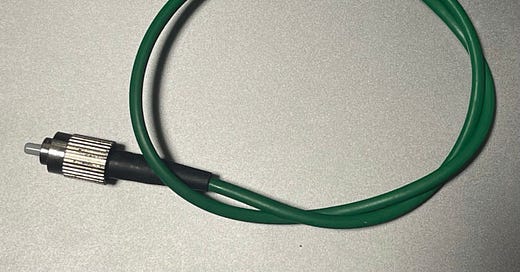
This fiber cable above comes from an Illumina Genome Analyzer. I’ve shown parts of this instrument in previous posts. It’s essentially reasonably cheap automated microscope, and as they can no longer be used for their intended purpose (DNA Sequencing) I’ve picked them up for as little as $61 on eBay.
The instrument also contains a Quantum GEM laser, which can be used for TIRF single molecule imaging. I’ve played with this and documented it on the blog.
This laser is fed into a multimode fiber, which is launched into a TIRF prism. I wanted to know what kind of fiber this was, so I could replace it, and buy compatible parts.
Multimode fibers are available with various different specifications, but one of the critical parameters is the core diameter, that is the size of the core that actually carries the light. And it this case, there were no markings indicating what it might be.
So I decided to slice one of the fibers open and take a look. In the diagram below you can see the general construction of the fiber cable:
The construction of these cables is surprisingly complex and a lot of it is really about protecting that tiny inner core.
In order to image the fiber itself and figure out how big the core was, I sliced off some of the inner fiber and stuck it to a slide with some JB Weld…
This let me take some reasonable pictures of the fiber under my epi-illuminated microscope at low magnification:
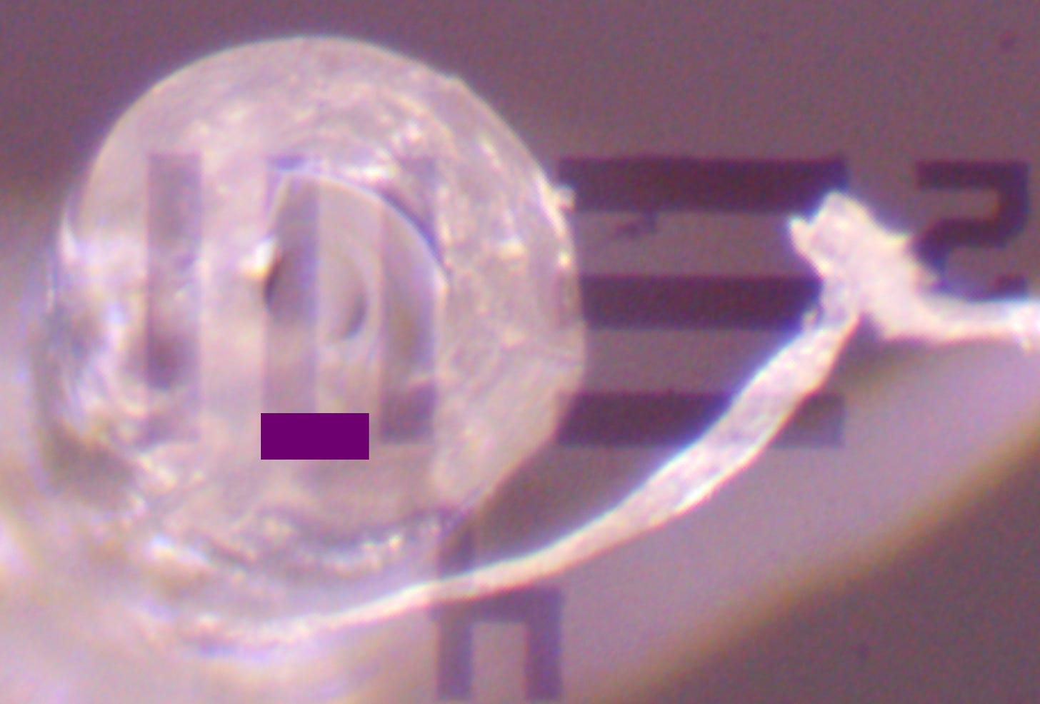
As a reference, I also took some snaps of a USAF1951 resolution target, and overlaid the images:
Lines on USAF1951 group 4 element 2 are 27.84 microns. By comparing the USAF1951 to the core I estimated a diameter of 50.112 microns.
Similarly, the outer cladding is 125.28 microns. Standard multimode fiber is either 50 or 62.5 micron with 125 micron cladding. So this suggests this fiber is 50/125 multimode fiber.
Yay! I can now buy replacement fiber, and other optical components for this rig…
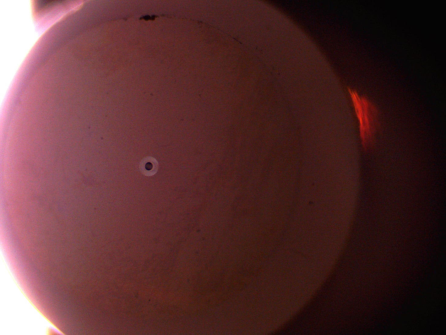
After doing all the above I decided to dermal off the end of the fiber connector itself and stick this under the microscope. Interestingly, you can similarly see the core and cladding. If I ever do this again, I will try and figure out how to non-destructively mount the fiber connector under a microscope.
Live and Learn!


